Clinical Translation of MRgHIFU

Clinical Translation of MRgHIFU

Clinical Translation of MRgHIFU

Clinical translation of MR-guided focused ultrasound for non-invasive liver tumor ablation.

Project Info

Start date

September 2024

End date

August 2028

Funding

Swiss National Science Foundation (SNSF) – Grant No. 10000596

Website

SNSF project page

Coordinator

HUG – Division of Radiology

Summary

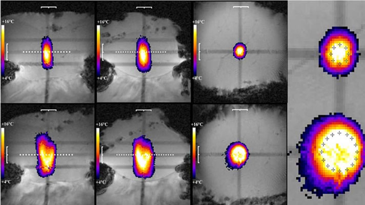
High-intensity focused ultrasound (HIFU) is a non-invasive technique for the thermal ablation of deep-seated tumors. Safe and effective treatment requires image guidance to plan the intervention and monitor the ablation process. Magnetic resonance imaging (MRI) is particularly well suited to this task because of its high soft-tissue contrast and its ability to monitor temperature changes during treatment, an approach known as MR-guided HIFU (MRgHIFU).

Treating abdominal organs such as the liver remains challenging because respiratory motion continuously changes the position of the target, introduces artifacts in MR thermometry, and can bring sensitive structures such as the thoracic cage or bowel into the ultrasound beam path.

The primary objective of this project is to conduct clinical trials of MRgHIFU for the treatment of neoplastic liver nodules. The project builds on a new MRgHIFU approach developed in a previous SNSF-funded research project, bringing the technology into clinical testing in patients for the first time. The studies progressively address the requirements for liver treatment, from accurate targeting to complete tumor ablation.

Within the project, Artanim contributes its expertise in computer science and real-time systems, including the design, implementation and quality assurance of software integrating self-scanning sonication with closed-loop temperature feedback control.

Partners

Rares Salomir – University Hospitals of Geneva, Division of Radiology
Imaging, data analysis, transducer development, clinical testing and project coordination.

Thibault Kössler, Alexis Ricoeur – University Hospitals of Geneva, Division of Oncology
Clinical trials and oncology expertise.

Andrea Peloso – University Hospitals of Geneva, Department of Surgery
Liver surgery.

Caecilia Charbonnier – Artanim
Design, implementation and quality assurance of real-time software integrating self-scanning sonication with closed-loop temperature feedback control.
Mocap RSA

Mocap RSA

Mocap RSA

A motion capture study of shoulder kinematics after reverse shoulder arthroplasty.

Project Info

Start date

January 2022

End date

June 2023

Funding

FORE Foundation / La Tour Hospital

Coordinator

Artanim

Summary

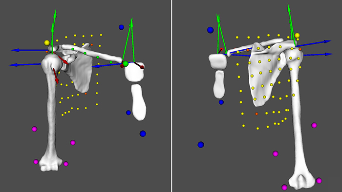
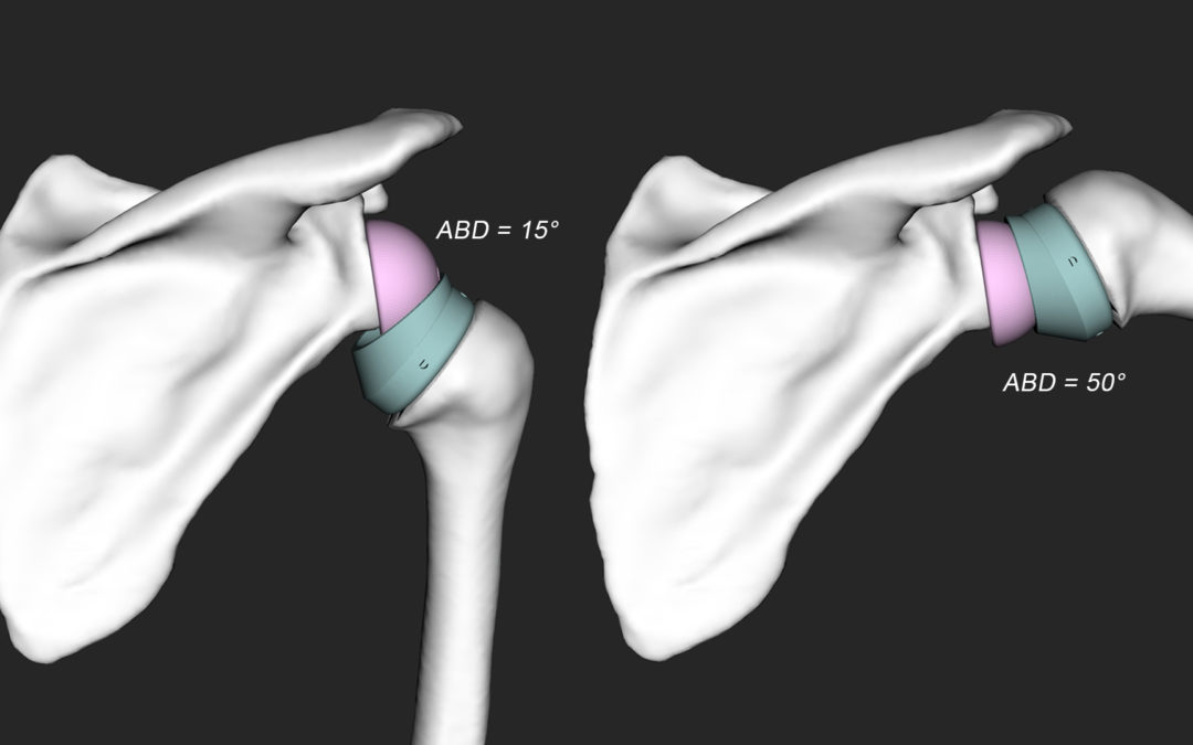
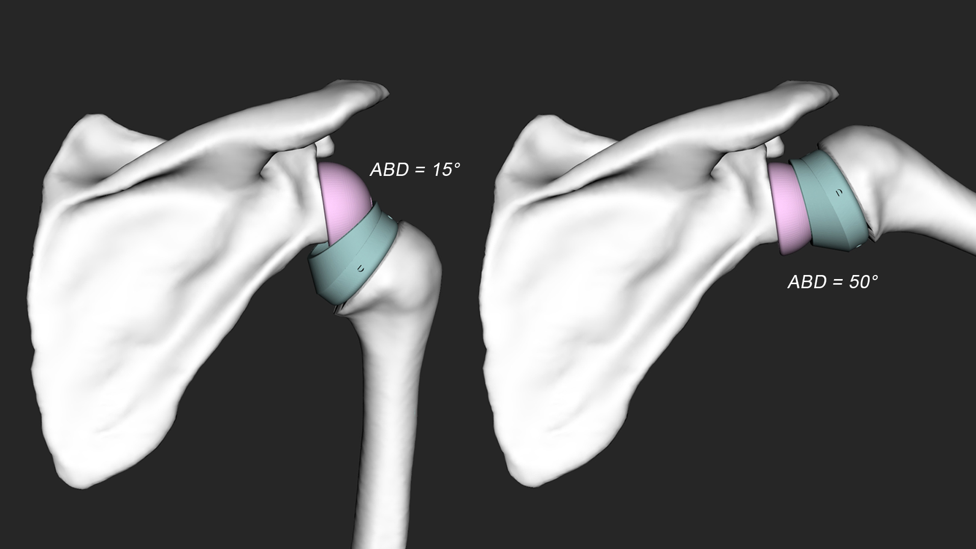
Mocap RSA investigates postoperative shoulder motion after reverse shoulder arthroplasty (RSA), with the aim of quantifying range of motion during activities of daily living and better understanding the resulting glenohumeral and scapulothoracic kinematics.

A major technical challenge is the accurate reconstruction of the postoperative anatomy and prosthetic components. Metallic implants can generate substantial artifacts in conventional CT imaging, making 3D reconstruction difficult. To address this, patients were scanned postoperatively using a dedicated low-dose cone-beam CT protocol, and a registration method was developed to align preoperative 3D bone models of the scapula and humerus, together with implant CAD models, onto the postoperative images.

Artanim was responsible for the segmentation and 3D reconstruction of the preoperative and postoperative bone and implant models, as well as for the dynamic simulation of reverse shoulder prostheses driven by motion capture data. The Haute École de Santé (HEdS) was responsible for the postoperative registration methodology, while La Tour Hospital handled patient recruitment and the clinical aspects of the study.

Partners

Artanim
Motion capture, 3D reconstruction and dynamic simulation of reverse shoulder prostheses.

Haute École de Santé (HEdS)
3D registration of postoperative RSA models.

La Tour Hospital – Elbow & Shoulder Orthopedics and Traumatology
Patient recruitment, clinical testing and medical supervision.

Related Publications

Levy G, Charbonnier C, Collin P, Pernoud A, Gaillard J, Lädermann A. Exploratory analysis of shoulder motion strategies for achieving functional hand behind the back in controls and after reverse shoulder arthroplasty. A study from the LaTour group, Int Orthop, 2026.
PDF · DOI

Levy G, Charbonnier C, Martinho T, Collin P, Bothorel H, Pernoud A, Lädermann A. Reverse Shoulder Arthroplasty: How to reach the back? A motion capture study to analyse glenohumeral strategies to perform functional internal rotation after RSA and in control cases, JSES Int, 10(2):101515, 2026.
PDF · DOI

Levy G, Charbonnier C, Martinho T, Collin P, Bothorel H, Pernoud A, Lädermann A. Analysis of the glenohumeral and scapula-thoracic motions ratio in controls and after reverse shoulder arthroplasty, Swiss Med Wkly, 2025.
PDF

Levy G, Charbonnier C, Martinho T, Collin P, Bothorel H, Pernoud A, Lädermann A. Reverse Shoulder Arthroplasty: How to reach the back? A motion capture study to analyse glenohumeral strategies to perform functional internal rotation after RSA and in control cases, 35th Annual Congress of the European Shoulder and Elbow Society (SECEC), Rotterdam, The Netherlands, September, 2025.
PDF

MRgHIFU Ablation of Liver Tumors

MRgHIFU Ablation of Liver Tumors

MRgHIFU Ablation of Liver Tumors

A research project advancing MR-guided focused ultrasound for non-invasive liver tumor ablation.

Project Info

Start date

July 2019

End date

December 2023

Funding

Swiss National Science Foundation (SNSF) – Grant No. 185308

Website

SNSF project page

Coordinator

HUG – Division of Radiology

Summary

High-intensity focused ultrasound (HIFU) is a non-invasive technique for the thermal ablation of deep-seated tumors. Safe and effective treatment requires image guidance to plan the intervention and monitor the ablation process. Magnetic resonance imaging (MRI) is particularly well suited to this task because of its high soft-tissue contrast and its ability to monitor temperature changes during treatment, an approach known as MR-guided HIFU (MRgHIFU).

Treating abdominal organs such as the liver remains challenging because respiratory motion continuously changes the position of the target, introduces artifacts in MR thermometry, and can bring sensitive structures such as the thoracic cage or bowel into the ultrasound beam path.

The goal of this project is to develop MRgHIFU methods for the treatment of unresectable liver malignancies using fast volumetric ablation. The research investigates an enlarged acoustic window, improved focusing capabilities, rapid automatic control of volumetric sonication, and self-scanning of the lesion by exploiting respiratory motion.

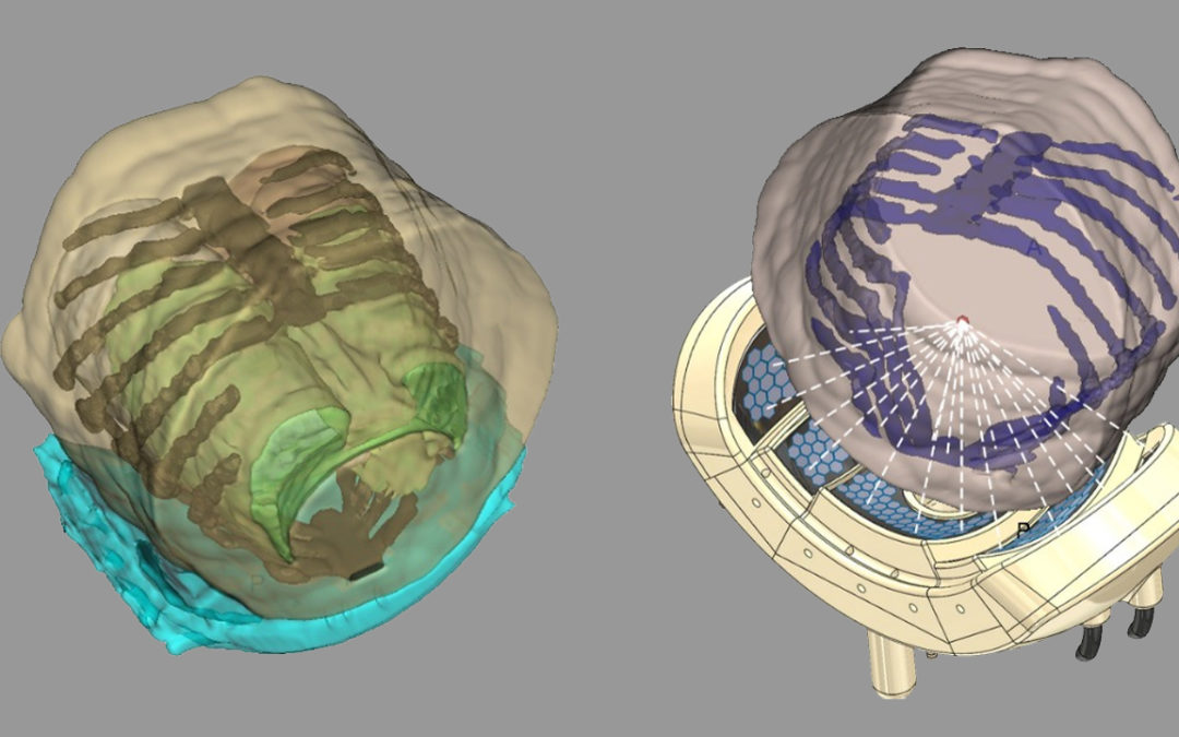
Within the project, Artanim was responsible for segmenting the target region and organs at risk, and for computing the optimal positioning of the robotic transducer system while taking into account the segmented anatomy and the required target coverage.

Partners

Rares Salomir, Sana Boudabbous, Alexis Ricoeur, Sylvain Terraz – University Hospitals of Geneva, Division of Radiology
Imaging, data analysis, transducer development, clinical testing and project coordination.

Christian Toso – University Hospitals of Geneva, Visceral Surgery
Liver surgery and clinical expertise.

Caecilia Charbonnier – Artanim
Segmentation of the target region and organs at risk, and optimization of robotic transducer positioning.

Related Publications

M’Rad Y, Charbonnier C, Elias de Oliveira M, Guillemin PC, Crowe LA, Kössler T, Poletti P-A, Boudabbous S, Ricoeur A, Salomir R, Lorton O. Computer-Aided Intra-Operatory Positioning of an MRgHIFU Applicator Dedicated to Abdominal Thermal Therapy Using Particle Swarm Optimization, IEEE Open J Eng Med Biol, 5:524–533, 2024.
PDF · DOI

Lorton O, Guillemin PC, M’Rad Y, Peloso A, Boudabbous S, Charbonnier C, Holman R, Crowe LA, Gui L, Poletti P-A, Ricoeur A, Terraz S, Salomir R. A Novel Concept of a Phased-Array HIFU Transducer Optimized for MR-Guided Hepatic Ablation: Embodiment and First In-Vivo Studies, Front Oncol, 12:899440, 2022.
PDF · DOI

Scapulo-Thoracic Alignment in B2 Glenoids

Scapulo-Thoracic Alignment in B2 Glenoids

Scapulo-Thoracic Alignment in B2 Glenoids

A 3D motion analysis study investigating scapulothoracic alignment in shoulder osteoarthritis.

Project Info

Start date

April 2019

End date

December 2019

Funding

FORE Foundation / La Tour Hospital

Coordinator

Artanim

Summary

Scapulo-Thoracic Alignment in B2 Glenoids investigates whether altered scapulothoracic alignment may contribute to the development of Walch type B2 glenoid osteoarthritis and its characteristic posterior erosion pattern.

The study uses a patient-specific 3D analysis combining medical imaging and optical motion capture to evaluate scapulothoracic alignment and compare pathological shoulders with the contralateral healthy side.

By characterizing the relationship between scapular orientation, glenohumeral mechanics and posterior glenoid erosion, the project aims to improve the understanding of shoulder pathoanatomy and the mechanisms involved in the development of osteoarthritis.

Within the project, Artanim was responsible for the dynamic simulation and analysis of shoulder kinematics using motion capture data.

Partners

Artanim
3D modeling and dynamic simulation of the shoulder.

La Tour Hospital – Elbow & Shoulder Orthopedics and Traumatology
Patient recruitment, clinical testing and medical supervision.

Imaging Center Rive Droite SA
Radiological imaging acquisitions.

Related Publications

Lädermann A, Athwal GS, Bothorel H, Collin P, Mazzolari A, Raiss P, Charbonnier C. Scapulothoracic Alignment Alterations in Patients with Walch Type B Osteoarthritis: An In Vivo Dynamic Analysis and Prospective Comparative Study, J Clin Med, 10(1):66, 2021.
PDF · DOI

Reverse Shoulder Arthroplasty

Reverse Shoulder Arthroplasty

Reverse Shoulder Prosthesis Simulation

A biomechanical simulation study evaluating implant configuration and shoulder range of motion.

Project Info

Start date

January 2017

End date

December 2017

Funding

Hirslanden Clinique La Colline

Coordinator

Artanim

Summary

Reverse Shoulder Prosthesis Simulation investigates how different implant and anatomical parameters affect shoulder range of motion after reverse shoulder arthroplasty (RSA).

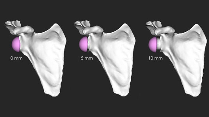
The study focuses in particular on glenoid lateralization, humeral inclination and critical shoulder angle (CSA), using computer-assisted simulations to evaluate their influence on joint mechanics and the risk of impingement or instability.

The objective is to identify surgical parameters that can help optimize implant configuration and reduce the risk of articular conflict and dislocation.

Within the project, Artanim was responsible for the dynamic simulation of reverse shoulder prostheses using kinematic data.

Partners

Artanim
Simulation of reverse shoulder prostheses.

Hirslanden Clinique La Colline – Center for Orthopaedic Surgery and Musculoskeletal Traumatology
Clinical testing and medical supervision.

Related Publications

Lädermann A, Tay E, Collin P, Piotton S, Chiu JCH, Michelet A, Charbonnier C. Effect of critical shoulder angle, glenoid lateralization and humeral inclination on range of motion in reverse shoulder arthroplasty, JSES Open Access, 3:240, 2019.
PDF · DOI

Lädermann A, Tay E, Collin P, Piotton S, Chiu JCH, Michelet A, Charbonnier C. Effect of critical shoulder angle, glenoid lateralization and humeral inclination on range of movement in reverse shoulder arthroplasty, Bone Joint Res, 8:378–386, 2019.
PDF · DOI

Charbonnier C, Tay E, Collin P, Piotton S, Chiu JCH, Michelet A, Lädermann A. Effect of critical shoulder angle, glenoid lateralization and humeral inclination on ROM in reverse shoulder arthroplasty, Swiss Med Wkly, 149(Suppl. 236):2S, 2019.
PDF