MRgHIFU Ablation of Liver Tumors

MRgHIFU Ablation of Liver Tumors

MRgHIFU Ablation of Liver Tumors

Minimally invasive intervention

Project Info

Start date:
July 2019

End date:
June 2023

Funding:
Swiss National Funds (SNF)

Coordinator:
HUG – Division of Radiology

Summary

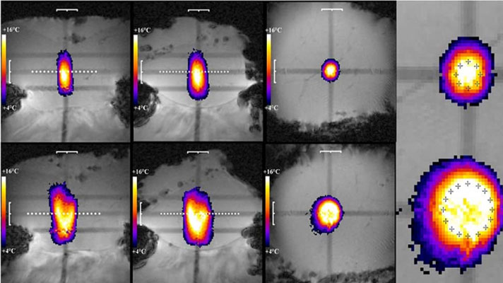
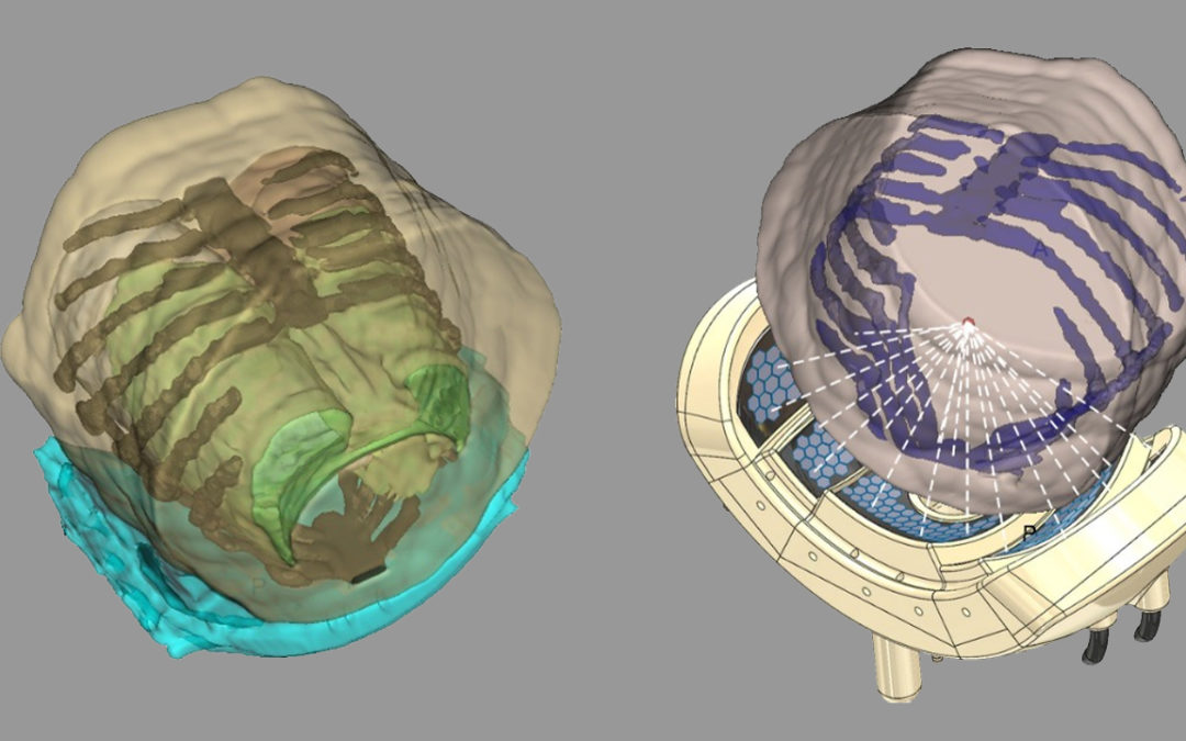
High intensity focused ultrasound (HIFU) is a precise method to thermally ablate deep-seated tumors in a non-invasive manner. A prerequisite for a safe and effective application of HIFU is image guidance, to plan and control the ablation process. The most suitable imaging modality is MRI, with its high soft tissue contrast and its ability to monitor tissue temperature changes (MR-guided HIFU, MRgHIFU). The therapy of abdominal organs, such as the liver, still poses several problems due to a moving target location caused by breathing, motion related MR-thermometry artefacts and near-field obstacles, i.e. thoracic cage or bowel.

The goal of this project is to treat unresectable liver malignancies with MRgHIFU, using fast volumetric ablation. Enlarged acoustic window and enhanced focusing number of the HIFU applicator, rapid automatic control of volumetric sonication, as well as self-scanning of the lesion by exploiting the respiratory motion are envisaged in this research.

In this project, Artanim was in charge of the segmentation of target region (tumor) and organs at risk, as well as the computation of the optimal transducer positioning of the robotic assistance system taking into account the segmented structures and the coverage target area.

Partners

University Hospitals of Geneva – Division of Radiology
Imaging, data analysis, transducer development, clinical tests and project coordination

Artanim
Segmentation of target region and organs at risk, transducer positioning

University Hospitals of Geneva – Division of Oncology
Clinical tests

Related Publications

Lorton O, Guillemin PC, M’Rad Y, Peloso A, Boudabbous S, Charbonnier C, Holman R, Crowe LA, Gui L, Poletti P-A, Ricoeur A, Terraz S, Salomir R. A Novel Concept of a Phased-Array HIFU Transducer Optimized for MR-Guided Hepatic Ablation: Embodiment and First In-Vivo Studies, Front Oncol, 30(12):899440, 2022.
PDF


Multiparametric Imaging of ORL Tumors

Multiparametric Imaging of ORL Tumors

Multiparametric Imaging of ORL Tumors

Contouring for radiotherapy planning

Project Info

Start date:
January 2018

End date:
December 2020

Funding:
Swiss National Funds (SNF)

Coordinator:
HUG – Division of Radiology

Summary

This project aimed to improve the image-based diagnosis and treatment of head and neck squamous cell carcinoma. Multiparametric imaging with morphological MRI, diffusion-weighted MRI (DW-MRI), dynamic contrast enhanced MRI (DCE-MRI) and PET may offer precise tumor and lymph node staging, improved risk stratification and effective management of patients with primary and recurrent head and neck cancer. Moreover, by combining multiparametric data, target volume delineation and biological dose painting for radiotherapy planning can be improved. The goal of this project was to improve tumor diagnosis and tailor therapeutic approaches in view of a personalized treatment strategy.

In this project, Artanim was in charge of 3D tumors reconstruction and participated in the development of an histologically validated computer-aided tumor model for the accurate contouring of gross tumor volume for radiotherapy planning.

Partners

University Hospitals of Geneva – Division of Radiology
Imaging, data analysis, radiologic-pathologic correlation and project coordination

Artanim
3D reconstruction and development of a tumor model

University Hospitals of Geneva – Division of Nuclear Medicine
Segmentation of PET images and methodological support

University of Geneva – Division of Clinical Pathology
Pathological evaluation and radiologic-pathologic correlation

University Hospitals of Geneva – Clinic of Otorhinolaryngology
Surgical dissection of tumor specimen and radiologic-pathologic correlation